Not an uncommon lesion, a nice example of a reticular network in a junctional naevus. Often patients ask to have these removed, replacing a benign lesion with a scar.
During a routine skin check, a mole was noticed that was larger and darker than the person’s other moles.
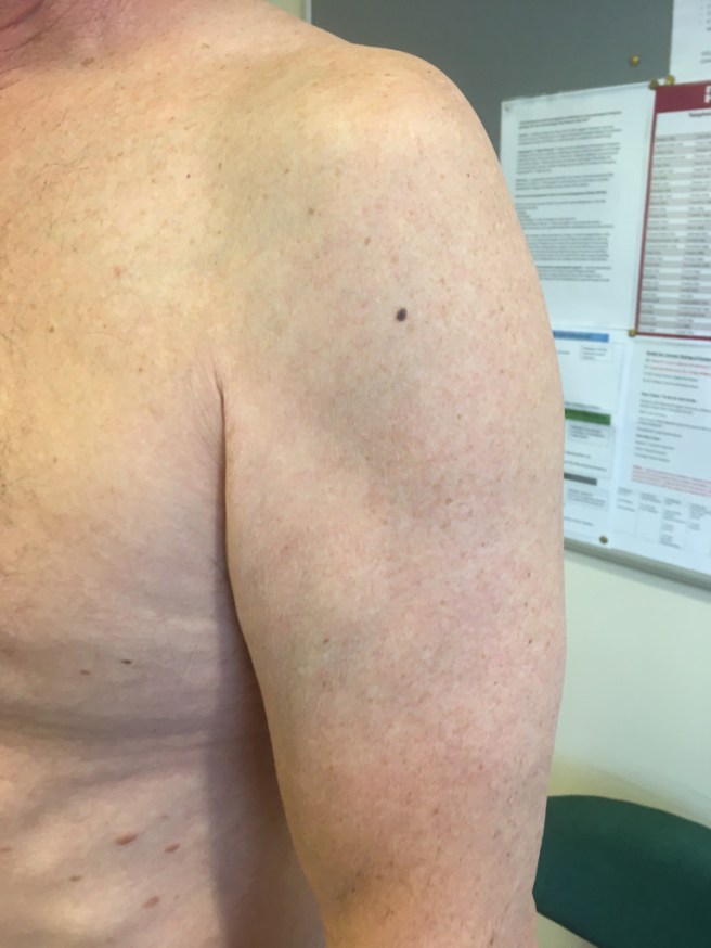
It’s not dramatic, but it stands out, so it needs a closer look.
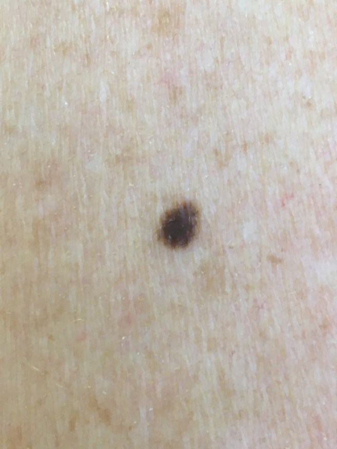
Dark, slightly irregular. Needs a closer look.
Dermoscopy reveals no chaos, clues to benignity, no melanoma clues.
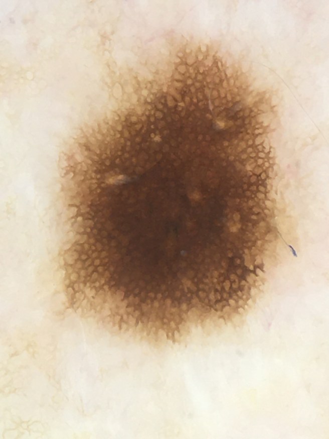
This mole is dark brown with two patterns arranged concentrically (featureless centre, reticular lines in the periphery which fade out evenly all round. We also see several small pale areas which represents perifollicular hypo pigmentation. This is of no significance. The overall appearance is highly symmetrical, a slight irregularity of shape means nothing.
Lesion was not excised. The beginner with Dermoscopy should get a book, attend courses and use the web, but should then apply the dermoscope to hundreds and thousands of lesions to ‘train your brain’ to recognize the range of normal naevi…
View original post 4 more words
